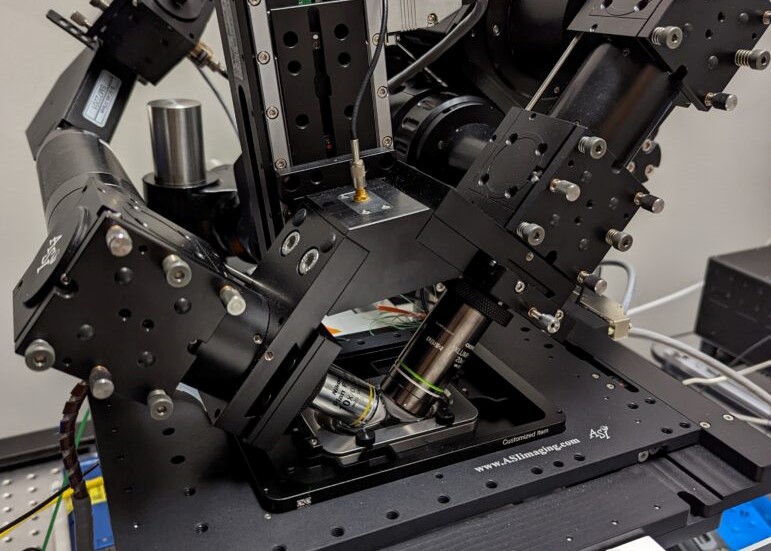
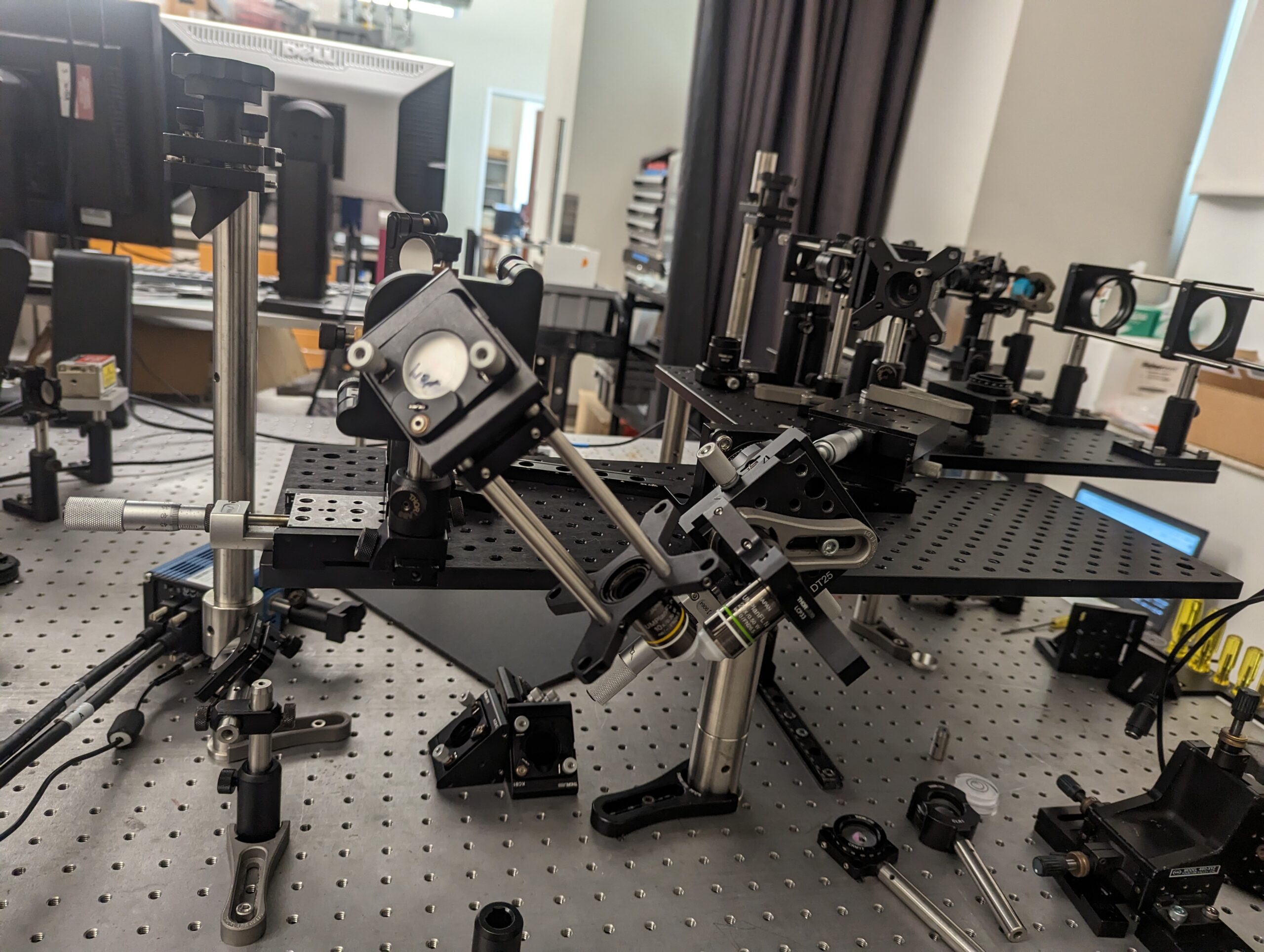
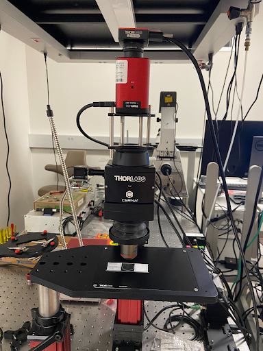
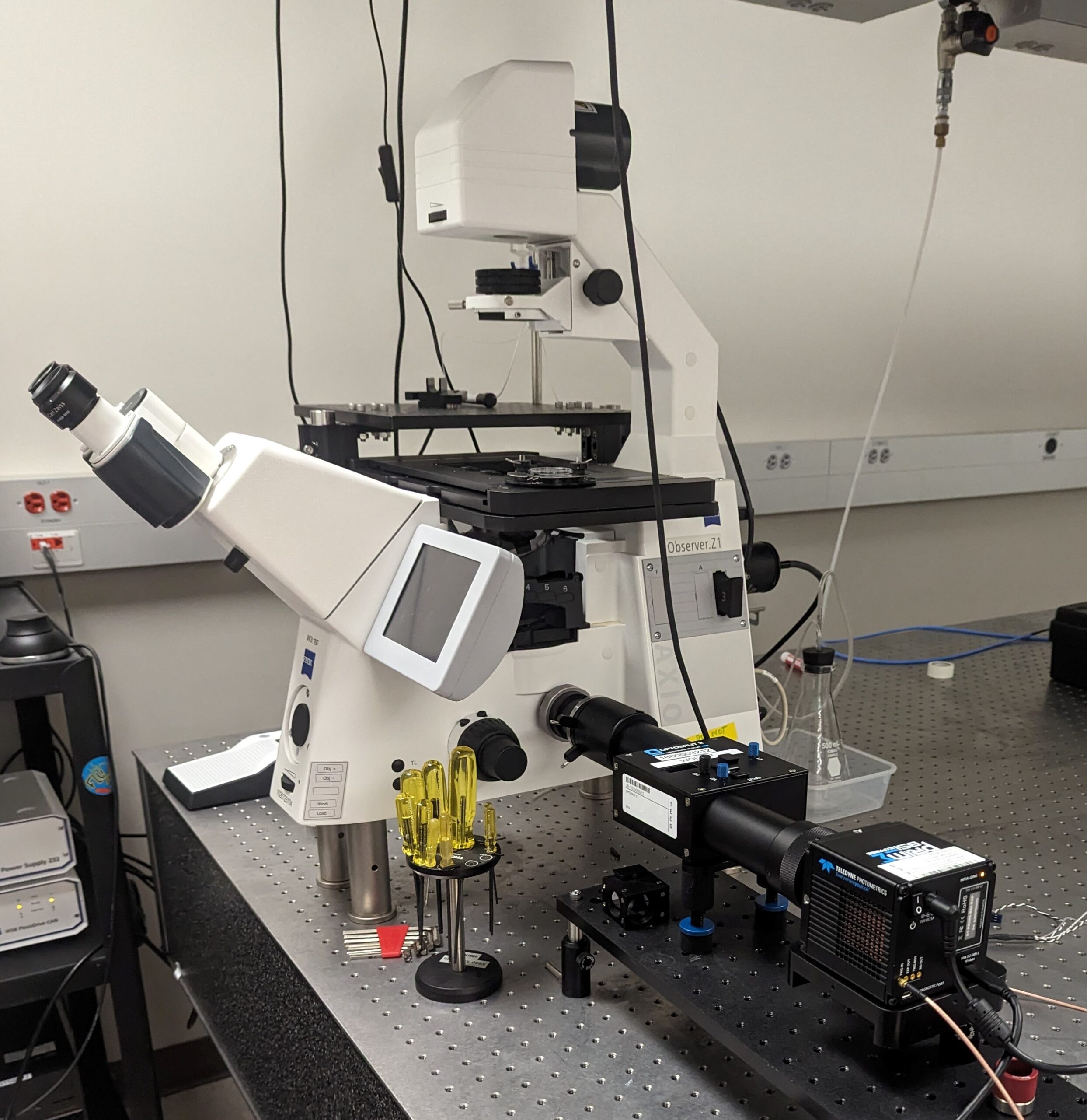
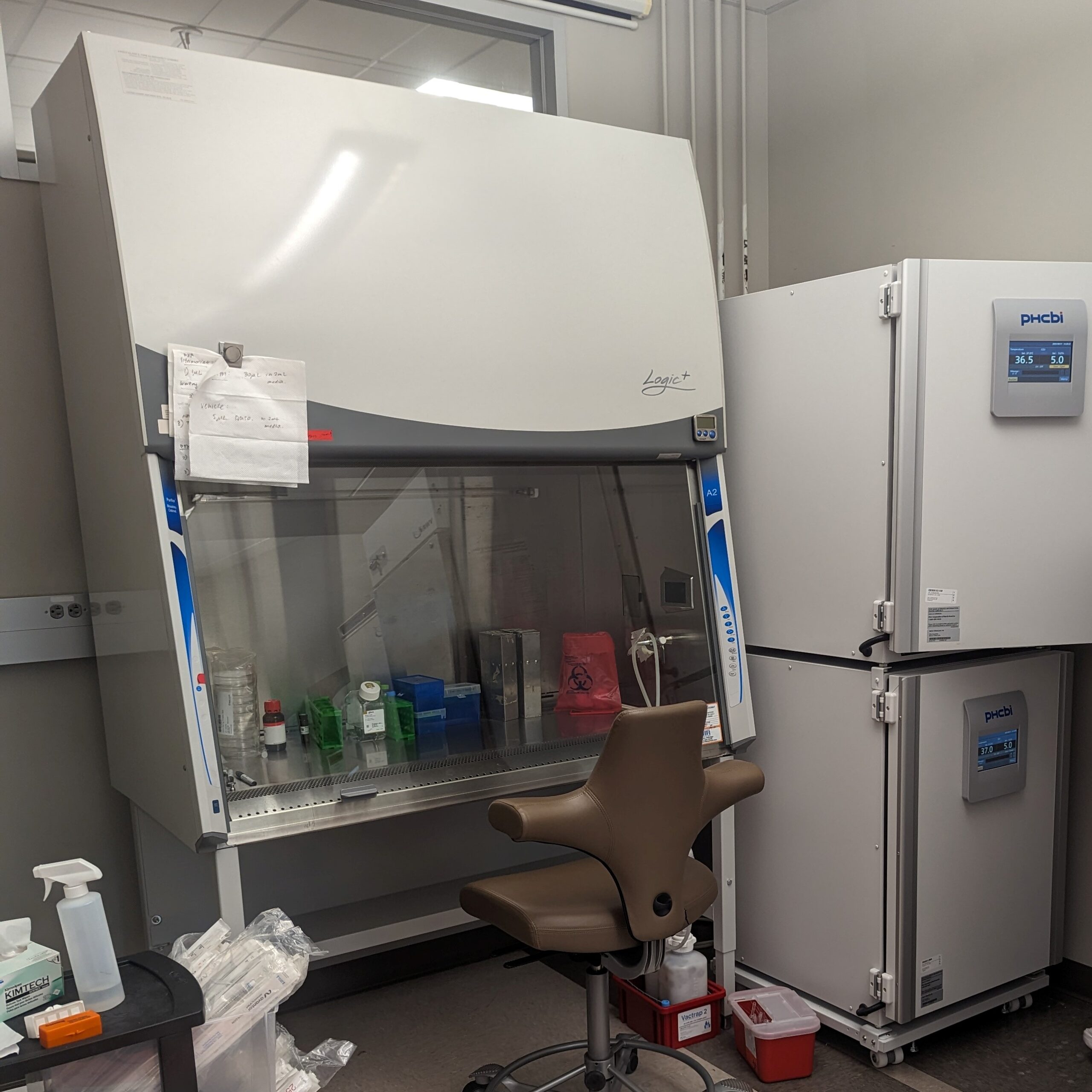
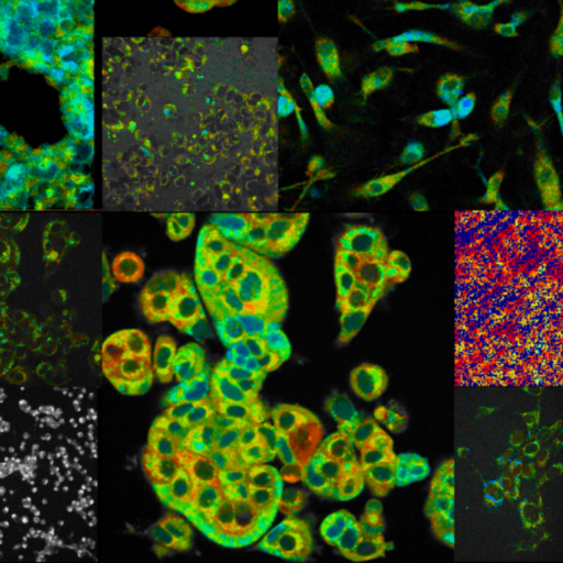
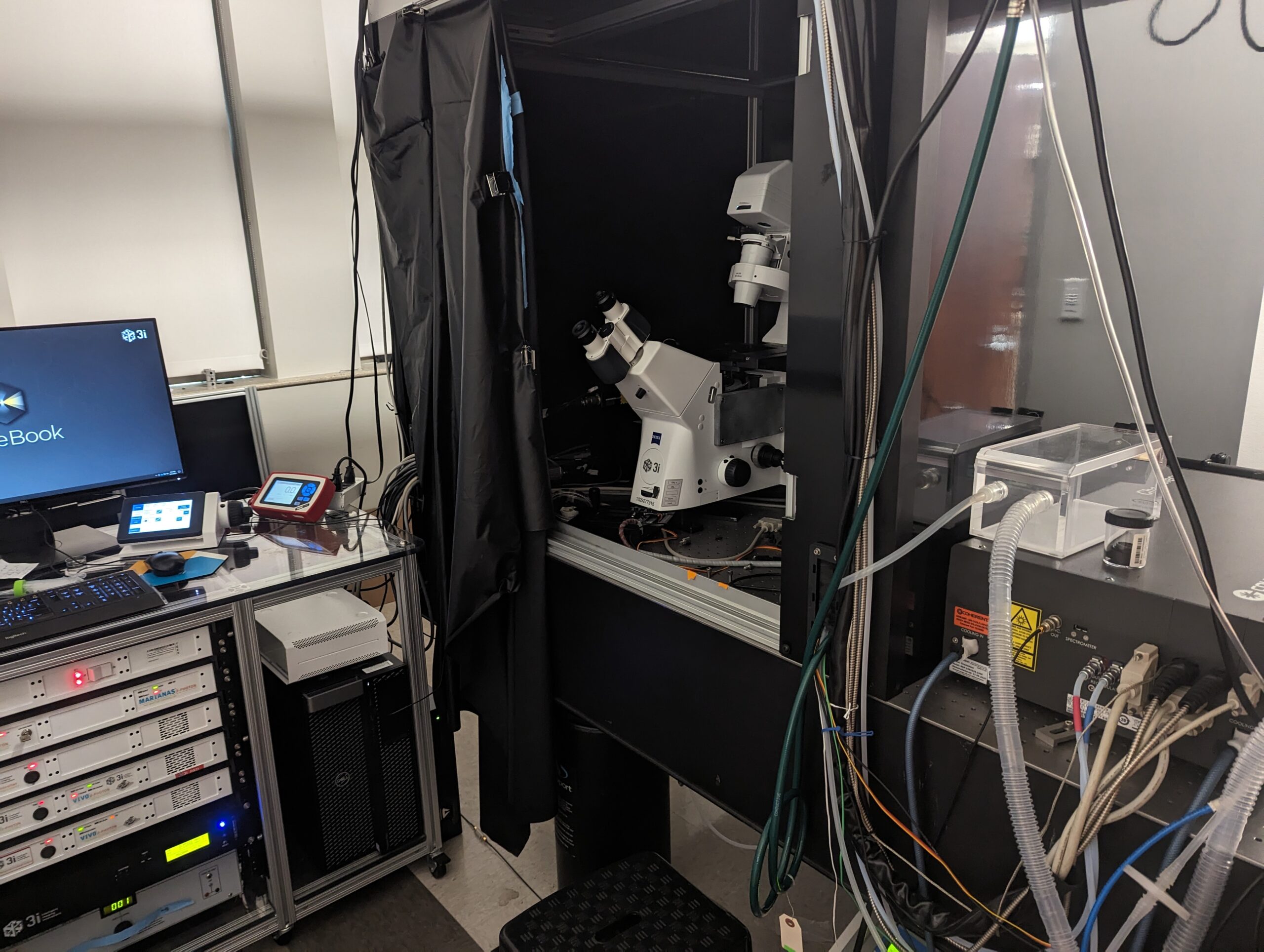
Multiphoton Fluorescence Lifetime Microscope

Multiphoton fluorescence lifetime microscope with tunable TiSapph laser (~680-1080 nm) for excitation. Two channel FLIM imaging plus a third intensity PMT. Inverted configuration with 20X, 0.8 NA air objective, 40X 1.1 NA water objective, and 100X 1.46 NA oil objective. Emission filters include 447/60 nm, 550/88 nm, and 675/67 nm. Environmental control for live cell and in vivo imaging.


Lightsheet with IR Stimulation

Lightsheet fluorescence microscope with integrated and co-aligned infrared laser. Lightsheet includes 375, 405, 488, 561, and 640 nm excitation lasers. A 10X 0.3 water dipping objective is used for illumination and a 20X water dipping objective is used for imaging. Two cameras allow separate, two color simultaneous imaging. Emission filters include 447/60, 525/50, 513/17, 550/88, 607/70, and 692/40 nm. Infrared light (1470 nm, 1940 nm) integrated into inverted objective (20X IR objective).


Hyperspectral System

Custom built diffuse reflectance and fluorescence hyperspectral microscope. Fluorescence excitation at 385 nm.


Autofluorescence Imaging

Inverted widefield fluorescence microscope with an image splitter for dual picture imaging on a single camera. Currently set up for simultaneous imaging of NADH and FAD. Excitation light provided by a white light LED (X-Cite) through a 357/44 nm filter. Image splitter contains a 496 nm dichroic and 415/106 nm and 560/94 nm emission filters. Objectives include 20X, 40X 1.2 NA, and 63X. Includes perfusion system for live neuron imaging.


Tissue Culture

Fully equipped tissue culture facility adjacent to the microscopy rooms.

Equipment may be shared with collaborators or rented through the ETB Cost Center.  Contact Dr. Walsh (walshaj [@] tamu.edu) if you are interested in any of our systems.

Software

Training

The Quantitative Optical Imaging Lab at Texas A&M University has unique expertise in fluorescence lifetime imaging, label-free optical imaging, customized optical system design, and advanced optical microscopy image analysis. We can provide training in these and related areas. Contact Dr. Walsh if you are interested in learning more.

Training Activities:

  • August 2023: Hosted visiting scientist from UT-Austin for a light-sheet microscope boot camp.  Over a week, covered optical system design and modeling in Optic Studio, built a double-objective light-sheet and built a single-objective light-sheet microscope.
  •  March 2023: Dr. Walsh served as a Faculty Instructor at the 20th Annual Workshop on FRET,  FLIM, and FLIRR (Metabolism)  Microscopy hosted by the W. M. Keck Center for Cellular Imaging (KCCI) University of Virginia, Charlottesville
  • Article and video on how to use fluorescence lifetime imaging of NADH and FAD to capture shifts in cellular metabolism (includes tips for setting up microscope excitation & emission filters, data analysis, and cyanide-perturbation protocol for testing/validation) – Autofluorescence Imaging to Evaluate Cellular Metabolism. Anna Theodossiou, Linghao Hu, Nianchao Wang, Uyen Nguyen, Alex J. Walsh, J. Vis. Exp. (177), e63282.
  • On-going: Multiphoton microscopy training of graduate students in collaborating labs.

Education

  • https://doi.org/10.1007/s43683-022-00070-7
    • Class activity on privilege.
    • Learning objectives for Justice, Equity, Diversity and Inclusion in Biomedical Engineering curriculum
    • “Design and Implementation of Privilege for Sale, a JEDI Activity for a Biomedical Engineering Introductory Course” Walsh, Raghavan, Biomedical Engineering Education 2022.
  •  Biophotonics Education …. coming soon