Recent Publications:
- Fluorescence intensity and lifetime redox ratios detect metabolic perturbations in T cells. Hu L, Wang N, Cardona E, and Walsh AJ, Biomedical Optics Express, 2020 (11)10: 5674-5688.
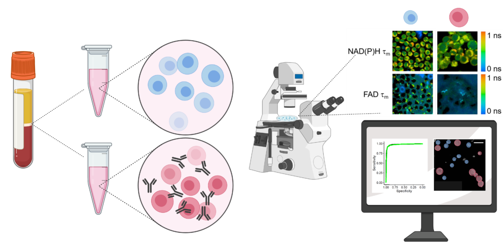
- Classification of T-cell activation via autofluorescence lifetime imaging. Walsh AJ*, Mueller KP, Tweed K, Jones I, Walsh CM, Piscopo NJ, Niemi NM, Pagliarini DJ, Saha K, Skala MC*, Nature Biomedical Engineering, 2020.
Currently, the activation of immune cells is assessed by label-based methods including flow cytometry and immunohistochemistry. Activated immune cells require high rates of glycolysis to maintain immune activities. Therefore, we are using fluorescence lifetime imaging of NAD(P)H and FAD which reports cellular metabolic information, to evaluate immune cell function. We have shown that the combination of machine learning classification of fluorescence imaging features achieves high accuracy (~98%) for classification of activation of T cells. Label-free imaging assays of immune cell function will allow unprecedented study of immune cell behaviors, functional assessment of immune cells in small volume samples (e.g. neonatal blood draws), and quality control/quality assurance of biomanufactured immune cells.